Abstract
Context: Hydrocephalus is the abnormal accumulation of cerebrospinal fluid (CSF) within the brain. The most common cause of hydrocephalus in premature babies is bleeding in the brain from an intraventricular hemorrhage. Intraventricular hemorrhage occurs in 15%–20% of infants less than 1500 g. The blood from an intraventricular hemorrhage blocks the absorption of CSF, causing it to build inside the ventricles. If severe, hydrocephalus can cause increased pressure inside the skull that can damage the brain if left untreated.
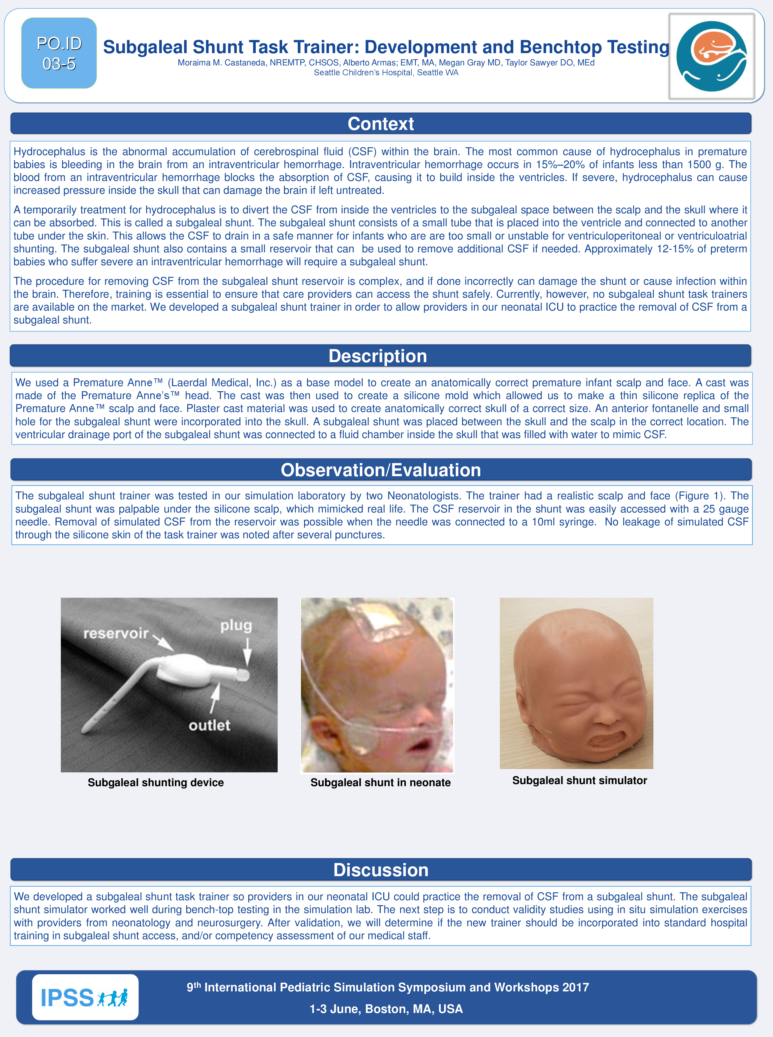
A temporarily treatment for hydrocephalus is to divert the CSF from inside the ventricles to the subgaleal space between the scalp and the skull where it can be absorbed. This is called a subgaleal shunt. The subgaleal shunt consists of a small tube that is placed into the ventricle and connected to another tube under the skin. This allows the CSF to drain in a safe manner for infants who are are too small or unstable for ventriculoperitoneal or ventriculoatrial shunting. The subgaleal shunt also contains a small reservoir that can be used to remove additional CSF if needed. Approximately 12-15% of preterm babies who suffer severe an intraventricular hemorrhage will require a subgaleal shunt.
The procedure for removing CSF from the subgaleal shunt reservoir is complex, and if done incorrectly can damage the shunt or cause infection within the brain. Therefore, training is essential to ensure that care providers can access the shunt safely. Currently, however, no subgaleal shunt task trainers are available on the market. We developed a subgaleal shunt trainer in order to allow providers in our neonatal ICU to practice the removal of CSF from a subgaleal shunt.
Description: We used a Premature Anne™ (Laerdal Medical, Inc.) as a base model to create an anatomically correct premature infant scalp and face. A cast was made of the Premature Anne’s™ head. The cast was then used to create a silicone mold which allowed us to make a thin silicone replica of the Premature Anne™ scalp and face. Plaster cast material was used to create anatomically correct skull of a correct size. An anterior fontanelle and small hole for the subgaleal shunt were incorporated into the skull. A subgaleal shunt was placed between the skull and the scalp in the correct location. The ventricular drainage port of the subgaleal shunt was connected to a fluid chamber inside the skull that was filled with water to mimic CSF.
Observation/Evaluation: The subgaleal shunt trainer was tested in our simulation laboratory by two Neonatologists. The trainer had a realistic scalp and face (Figure 1). The subgaleal shunt was palpable under the silicone scalp, which mimicked real life. The CSF reservoir in the shunt was easily accessed with a 25 gauge needle. Removal of simulated CSF from the reservoir was possible when the needle was connected to a 10ml syringe. No leakage of simulated CSF through the silicone skin of the task trainer was noted after several punctures.
Discussion: We developed a subgaleal shunt task trainer so providers in our neonatal ICU could practice the removal of CSF from a subgaleal shunt. The subgaleal shunt simulator worked well during bench-top testing in the simulation lab. The next step is to conduct validity studies using in situ simulation exercises with providers from neonatology and neurosurgery. After validation, we will determine if the new trainer should be incorporated into standard hospital training in subgaleal shunt access, and/or competency assessment of our medical staff.