Selective Retinal Therapy with Microsecond Exposures Using a Continuous Line Scanning Laser
Abstract
Purpose: To evaluate the safety, selectivity, and healing of retinal lesions created using a continuous line scanning laser.
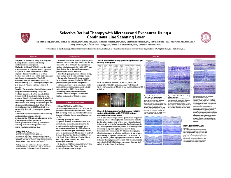
Methods: A 532 nm Nd:YAG laser with retinal beam diameters of 40 and 66 μm was applied to 60 eyes of 30 Dutch-Belted rabbits. Retinal exposure duration varied from 15 to 60 μs. Lesions were acutely assessed by ophthalmoscopy and fluorescein angiography (FA). RPE flatmounts were evaluated with LIVE/DEAD fluorescent assay (LD). Histological analysis was performed at 7 time points from 1 hour to 2 months.
Results: The ratios of the threshold of rupture and of ophthalmoscopic visibility (OV) to FA visibility (measures of safety and selectivity) increased with decreasing duration and beam diameter. FA and LD yielded similar thresholds of RPE damage. Above the OV threshold, histology showed focal RPE damage and photoreceptor loss at one day, without inner retinal effects. By one week, photoreceptor and RPE continuity was restored. By 1 month, photoreceptors appeared normal.
Conclusion: Retinal therapy with a fast scanning continuous laser achieves selective
treatment of the RPE and, at higher power, of the photoreceptors without permanent
scarring or inner retinal damage. Continuous scanning laser can treat large retinal areas
within standard eye fixation time.
Related articles