Concordance of ECoG Mapping and rs-fMRI Mapping in Simple Motor Tasks
Abstract
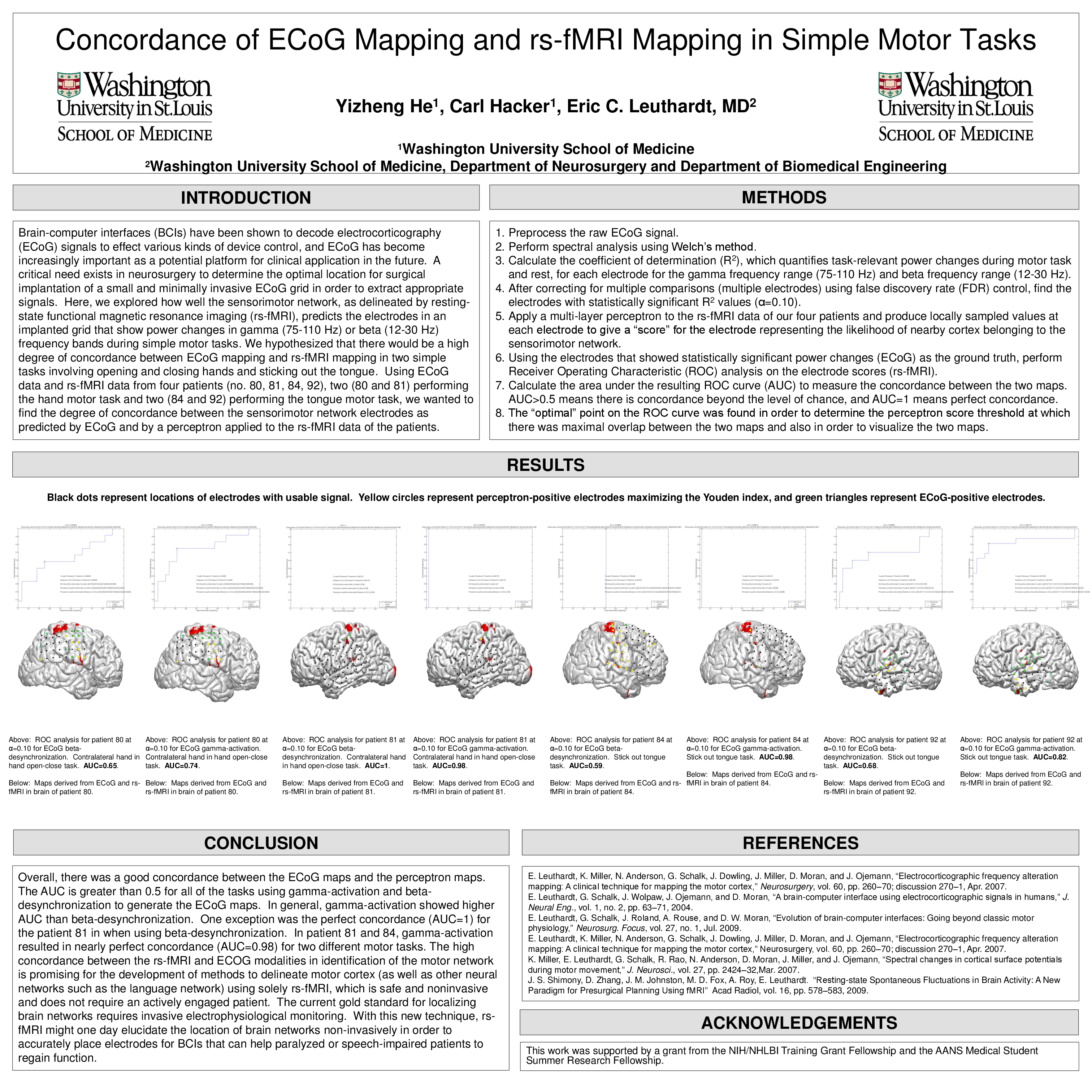
Brain-computer interfaces (BCIs) have been shown to decode electrocorticography (ECoG) signals to effect various kinds of device control, and ECoG has become increasingly important as a potential platform for clinical application in the future. A critical need exists in neurosurgery to determine the optimal location for surgical implantation of a small and minimally invasive ECoG grid in order to extract appropriate signals. Here, we explored how well the sensorimotor network, as delineated by resting-state functional magnetic resonance imaging (rs-fMRI), predicts the electrodes in an implanted grid that show power changes in gamma (75-110 Hz) or beta (12-30 Hz) frequency bands during simple motor tasks. We hypothesized that there would be a high degree of concordance between ECoG mapping and rs-fMRI mapping in two simple tasks involving opening and closing hands and sticking out the tongue. Using ECoG data and rs-fMRI data from four patients (no. 80, 81, 84, 92), two (80 and 81) performing the hand motor task and two (84 and 92) performing the tongue motor task, we wanted to find the degree of concordance between the sensorimotor network electrodes as predicted by ECoG and by a perceptron applied to the rs-fMRI data of the patients.
Overall, there was a good concordance between the ECoG maps and the perceptron maps. The AUC is greater than 0.5 for all of the tasks using gamma-activation and beta-desynchronization to generate the ECoG maps. In general, gamma-activation showed higher AUC than beta-desynchronization. One exception was the perfect concordance (AUC=1) for the patient 81 in when using beta-desynchronization. In patient 81 and 84, gamma-activation resulted in nearly perfect concordance (AUC=0.98) for two different motor tasks. The high concordance between the rs-fMRI and ECOG modalities in identification of the motor network is promising for the development of methods to delineate motor cortex (as well as other neural networks such as the language network) using solely rs-fMRI, which is safe and noninvasive and does not require an actively engaged patient. The current gold standard for localizing brain networks requires invasive electrophysiological monitoring. With this new technique, rs-fMRI might one day elucidate the location of brain networks non-invasively in order to accurately place electrodes for BCIs that can help paralyzed or speech-impaired patients to regain function.
Related articles