Keratoconus Diagnostic Model Integrating Ocular Response Analyzer Measurements and Corneal Topography
Abstract
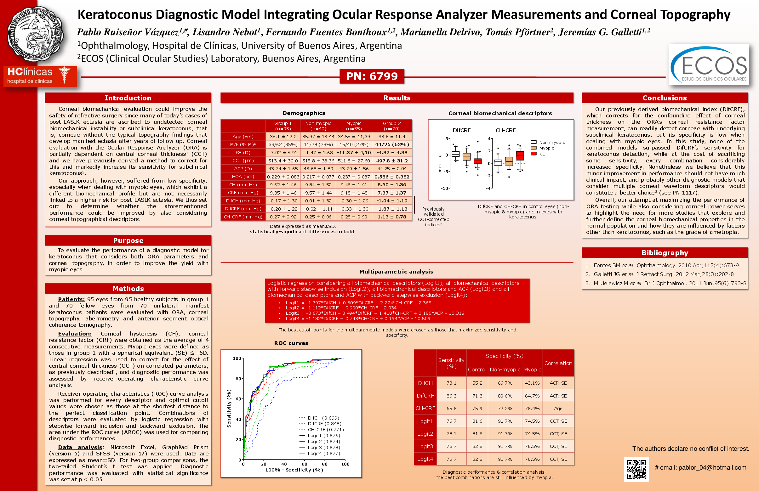
Purpose: To evaluate the performance of a diagnostic model for keratoconus that considers both Ocular Response Analyzer (ORA) parameters and corneal topography, in order to improve the yield with myopic eyes.
Methods: 95 eyes from 95 healthy subjects in group 1 and 70 eyes from 70 keratoconus patients in group 2 were evaluated with Ocular Response Analyzer (ORA), corneal topography, aberrometry and anterior segment optical coherence tomography for central corneal thickness (CCT) measurement. For group 1, an eye with spherical equivalente < -5.00 was considered myopic and for group 2, the eye with the lowest average corneal power (ACP) was chosen. Corneal hysteresis (CH) and resistance factor (CRF) transformations to compensate for CCT effect (DifCH, DifCRF and CH-CRF) and diagnostic cutoff points were obtained from previous work on an independent sample. Discriminant functions were built using biomechanical variables and ACP.
Results: Group 2 eyes had lower CCT (μm, 516.6±35.87 vs 491.6±31.92, p<0.01), DifCH (-0.121±1.35 vs -1.01±1.295, p<0.01), DifCRF (-0.147±1.50 vs 1.774±1.468, p<0.01) and higher CH-CRF (0.25±0.92 vs 1.1±0.96, p<0.01) and average corneal power (ACP, 43.96±1.780 vs 45.99±4.248, p<0.01). 40 eyes in group 1 were non-myopic and 55 eyes were myopic. The DifCRF cutoff point correctly diagnosed 67.5% of group 1 eyes (77.5% of non-myopic and 60.0% of myopic eyes) and 84.3% of group 2 eyes, whereas the best discriminant function (combining DifCRF, DifCH, CH-CRF and ACP) correctly identified 84.2% of group 1 eyes (92.5% of non-myopic and 78.2% of myopic eyes) and 75.7% of group 2 eyes.
Conclusions: DifCRF readily detects subclinical biomechanical abnormalities of keratoconic corneas (high sensitivity) but falsely flags biomechanically atypical, normal myopic eyes (reduced specificity). An integrated approach combining corneal biomechanics and anterior curvature increases the model’s specificity with some sacrifice in sensitivity, probably improving its usability in the preoperative evaluation of refractive surgery candidates.
Related articles