Heart Rate Variability Monitoring Following Brain Trauma
Abstract
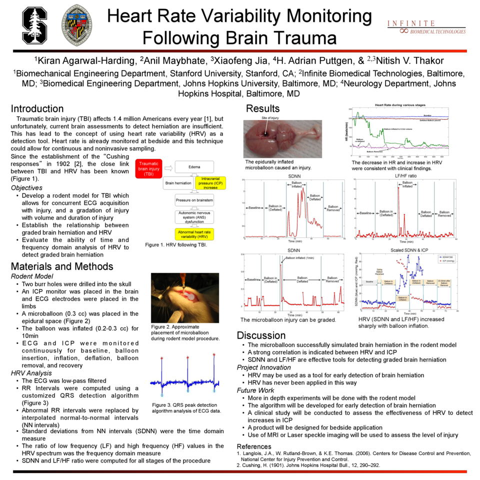
Traumatic brain injury (TBI) affects 1.4 million Americans every year. Heightened intracranial pressure (ICP) and subsequent brain herniation is a commonly observed event among those who clinically deteriorate after TBI. Prompt detection of brain herniation is important to prevent further injury. Heart rate variability (HRV) may provide an easy, noninvasive tool for evaluating TBI patients and detecting early onset of herniation. Normal heart rate is regulated by the autonomic nervous system (ANS), which is centrally regulated by the hypothalamus and brainstem. During herniation, the brainstem is compressed, causing ANS dysfunction and eventual abnormal HRV.
We have developed a rodent model to analyze the relationship between HRV and graded brain herniation by progressively inflating an intracranial balloon while simultaneously recording surface ECG and ICP signals. The model was successful in simulating graded brain herniation and gave consistent, reproducible results. Both the time domain analysis and frequency domain analysis were found to be effective in detecting the injury. HRV was observed to increase only with balloon inflation, with no false positive results.
This indicates that HRV can be used as an effective tool for detecting brain herniation in the clinical setting. Further research will be done on the rodent model and eventually the algorithm will be adapted to detect brain herniation. The algorithm will be studied in a clinical setting, leading to the development of a bedside device for HRV monitoring and early detection of brain herniation. This will lead to significant improvements in patient care by allowing prompt intervention, thereby preventing irreversible coma and death.
Related articles