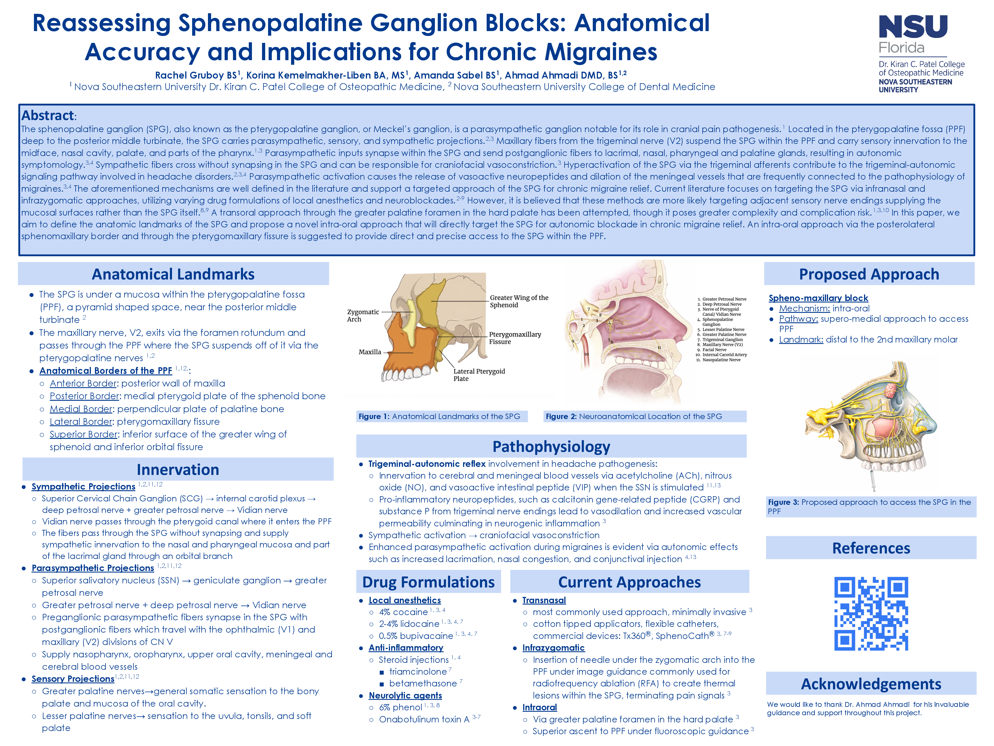
Abstract
Background: The sphenopalatine ganglion (SPG), also known as the pterygopalatine ganglion, is a parasympathetic ganglion notable for its role in cranial pain pathogenesis. Located in the pterygopalatine fossa (PPF), the SPG carries parasympathetic, sensory, and sympathetic projections. Hyperactivation of the SPG via trigeminal afferents contributes to the trigeminal-autonomic signaling pathway implicated in chronic migraine pathophysiology. SPG blockade techniques exist as a therapeutic option for intractable migraine relief; however, the anatomic accuracy of targeting the SPG remains uncertain.
Methods: A focused narrative literature review was conducted to evaluate SPG anatomy, pathophysiology, and current practices of SPG blockades for chronic migraine relief. Literature searches were performed using PubMed. The following key words were used in our primary search: “sphenopalatine ganglion block”, “pterygopalatine ganglion”, “chronic migraine”, and “refractory migraine”. Articles published within the last 10 years that discussed techniques to SPG blocks, namely: infranasal, infrazygomatic, and transoral approaches, as well as varying anesthetic and neuromuscular blocking agents were included. Articles discussing SPG blockade in the setting of other non-migraine relief were excluded from the primary search.
Results: The sphenopalatine ganglion’s anatomical location and connections to autonomic and sensory projections to the face and cranium make it a viable target for therapeutic interventions in chronic migraine relief. SPG activation releases vasoactive neuropeptides that dilate meningeal vessels involved in migraines. Current literature focuses on blocking the SPG via infranasal or infrazygomatic approaches, utilizing varying drug formulations of local anesthetics and neuroblockades. A transoral approach through the greater palatine foramen of the hard palate has been attempted, though it poses greater complexity and complication risk. Upon anatomical analysis, it is believed that these methods are more likely targeting the distal nerve endings that project from the SPG, rather than the ganglion itself. Because of the inconsistency within current SPG blockade protocols, there remains a need for a more precise and anatomically accurate technique to directly target the ganglion.
Conclusion: Current practices of SPG blockade for chronic migraine relief may not consistently achieve direct ganglionic blockade within the PPF. This review aims to highlight the need for an anatomically accurate approach that will more reliably target the SPG. We propose an intra-oral approach directed supero-medially, distal to the second maxillary molar. This trajectory allows for access into the PPF via the posterolateral sphenomaxillary border at the pterygomaxillary fissure. This approach may offer improved specificity for autonomic blockade and represents a potential advancement in interventional management of chronic migraine.