Cardiomyocytes Generate Cardiomyocytes During Development, Aging, And Myocardial Infarction In Mice
Abstract
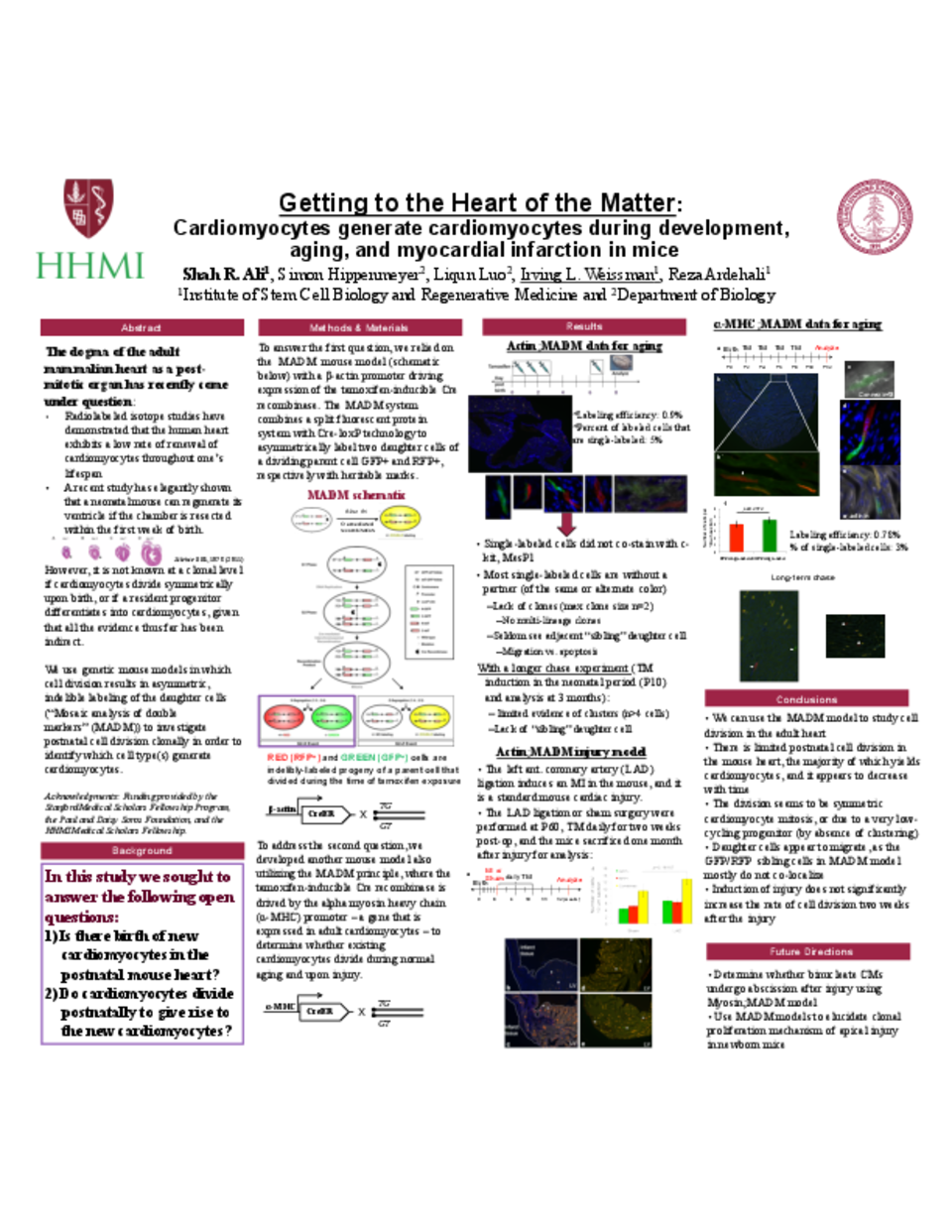
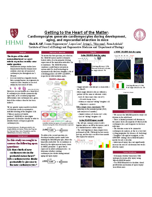
The mammalian heart has long been considered a post-mitotic organ, with conventional dogma maintaining that the total number of cardiomyocytes is set at birth. Analysis of cell division in the mammalian heart is complicated by cardiomyocyte binucleation shortly after birth, which makes it challenging to interpret traditional assays of cell turnover that rely on proxies of cell division. Recently the low rate of renewal of cardiomyocytes in humans after birth was elegantly calculated by measuring nuclear 14C content in the heart, providing the strongest evidence to date of this phenomenon3. Despite this advance, the nature of postnatal cardiomyogenesis remains poorly understood at the cellular level. Here, we extend upon these previous findings to show that the cell of origin of postnatal cardiomyogenesis is a differentiated alpha-myosin heavy chain (Myh6)-expressing cardiomyocyte using the “mosaic analysis with double markers” (MADM) mouse model4. Our results are consistent with life-long symmetric division of cardiomyocytes as a rare event that is evident in utero but significantly tapers after the first month of life in mice. Further, after ligation of the left anterior descending coronary artery that leads to a myocardial infarction in the MADM mice, we do not find an increase in the rate of cardiomyocyte division above the basal level in up to two weeks after the injury. The clonal analysis described herein is the first direct evidence of postnatal mammalian cardiomyogenesis.
Related articles