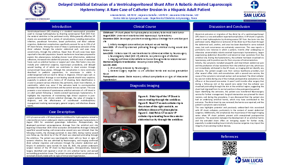
Abstract
Ventriculoperitoneal (VP) shunt complications are well documented, but spontaneous extrusion
through the umbilicus is exceedingly rare in adults, with fewer than 30 reported cases. Risk factors
include previous abdominal surgery, local inflammation, and adhesion-related pressure. Early
recognition is vital to prevent infection and facilitate safe removal. A 35-year-old woman with a VP
shunt placed in childhood for a dermoid brain tumor underwent robotic-assisted laparoscopic
hysterectomy in August 2024. She subsequently developed persistent serosanguineous drainage
from the umbilical port site starting in December 2024, initially attributed to superficial wound
healing. In June 2025, extrusion of the distal VP shunt tip through the umbilicus was observed
during Wound Care follow-up. The patient was admitted by Neurosurgery. She was neurologically
intact (GCS 15) and without systemic signs of infection. Imaging confirmed distal catheter
migration and extrusion through the anterior abdominal wall. She was started on empiric IV
Vancomycin and Cefepime. On June 30, 2025, she underwent VP shunt removal by Neurosurgery.
Intraoperatively, General Surgery identified and repaired a small (<1 cm) umbilical hernia and
excised surrounding granulation tissue without complications. Postoperatively, the patient
remained clinically stable with no signs of intracranial hypertension. This case illustrates the
importance of maintaining high clinical suspicion for rare VP shunt complications in postlaparoscopic
patients presenting with chronic wound drainage. A multidisciplinary approach,
including imaging, infectious prophylaxis, and combined neurosurgical and general surgical
intervention, was critical to successful resolution. Reporting such cases expands awareness of this
unusual complication and its management.


