Abstract
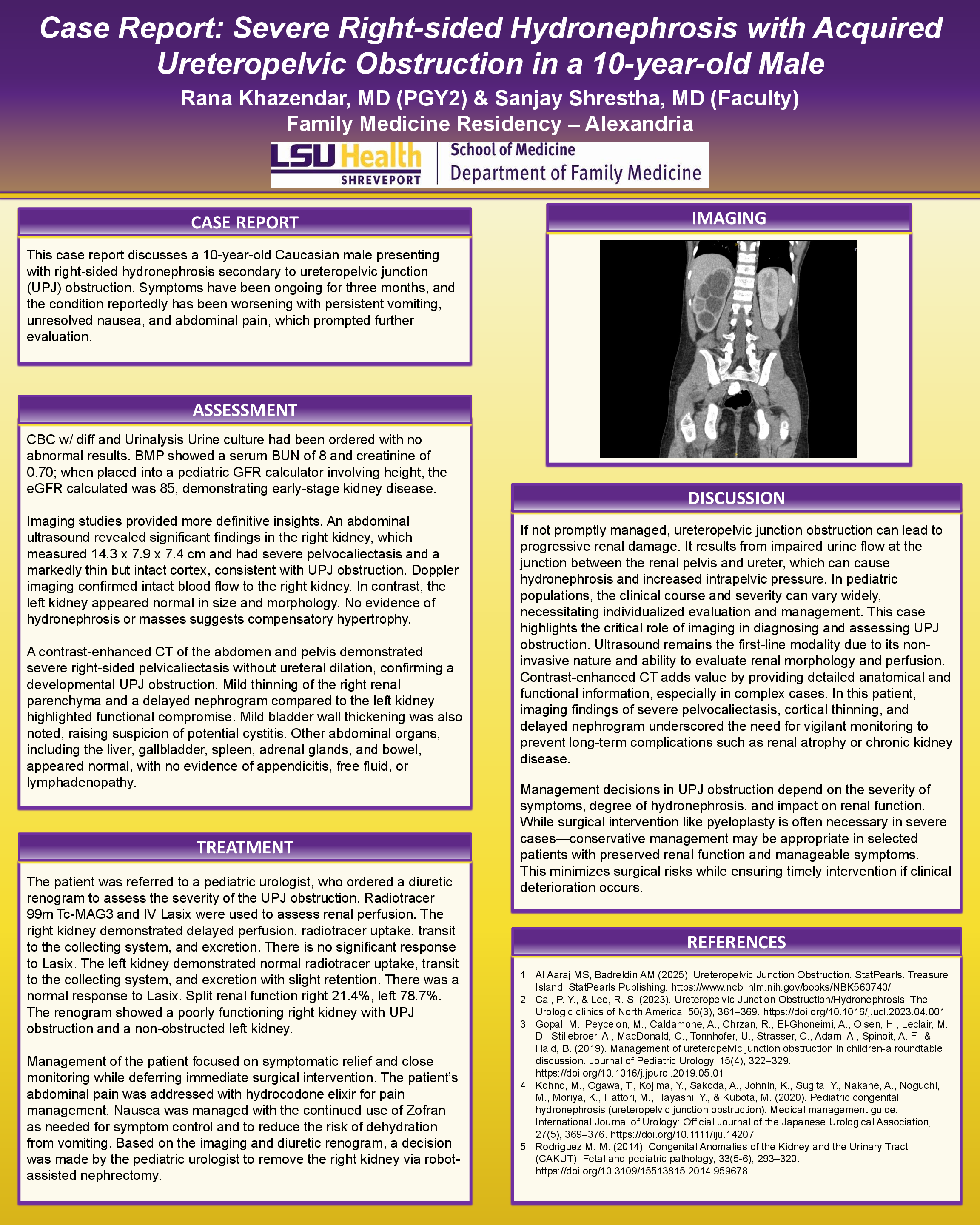
If not promptly managed, ureteropelvic junction obstruction can lead to progressive renal damage. It results from impaired urine flow at the junction between the renal pelvis and ureter, which can cause hydronephrosis and increased intrapelvic pressure. In pediatric populations, the clinical course and severity can vary widely, necessitating individualized evaluation and management. This case highlights the critical role of imaging in diagnosing and assessing UPJ obstruction. Ultrasound remains the first-line modality due to its noninvasive nature and ability to evaluate renal morphology and perfusion. Contrast-enhanced CT adds value by providing detailed anatomical and functional information, especially in complex cases. In this patient, imaging findings of severe pelvocaliectasis, cortical thinning, and delayed nephrogram underscored the need for vigilant monitoring to prevent long-term complications such as renal atrophy or chronic kidney disease.