Abstract
Objectives:
Patients with High grade glioma (HGG) who underwent chemoradiation usually have routine Magnetic
Resonance Imaging (MRI) every 2-3 months for surveillance. Quite often, the treatment related changes
including radiation necrosis (RN) and pseudo-progression (PP) are indistinguishable from tumor
recurrence (TR), which can cause significant dilemma to the treating physician. The main aim of this
study is to investigate the utility of Fluoro-dihydroxyphenylalanine Positron Emission Tomography ( 18 F -
DOPA PET) scan mapped with radiotherapy planning dosimetry in differentiating true progression from
RN and PP during follow up, and the potential application in further treatment management.
Methods and Materials:
We retrospectively reviewed ten accrued HGG patients over a duration of 12 months, who were initially
treated with concurrent chemoradiation and found to have clinical and/or MRI changes suggesting TR or
RN on follow up. Each patient underwent a 18 F-DOPA PET scan and the images were mapped with the
corresponding CT and MRI radiotherapy plan. We then analyzed the overlap of fused images with the
contours of primary tumor and target volumes. Descriptive analysis was used to identify TR and RN, and
patterns of correlation with dosimetry.
Results:
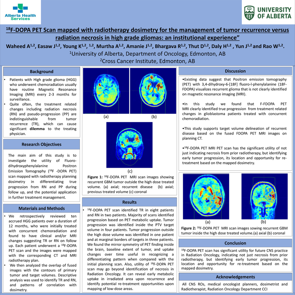
18 F-DOPA PET scan identified TR in eight patients and RN in two patients. Majority of scans identified
progression based on PET metabolic uptake. Tumor progression was identified inside the PTV target
volume in four patients. Tumor progression outside the high dose volume was identified in one patient
and at marginal borders of targets in three patients. We found the mirror symmetry of PET finding inside
the brain, baseline extent of tumor, and uptake changes over time useful in recognizing a differentiating
pattern when compared with the initial planning scan. Also, utility of 18 F-DOPA PET scan may go beyond
identification of necrosis in Radiation Oncology. It can reveal early metabolic uptake in irradiated area
upon recurrence and identify potential re-treatment opportunities upon mapping of low-dose areas.
Conclusion:
18 F-DOPA PET scan has significant utility for future CNS practice in Radiation Oncology, indicating not just
necrosis from prior radiotherapy, but identifying early tumor progression, its location and opportunity
for re-treatment based on the mapped dosimetry.


