Abstract
Purpose:
The differences between prostate CT contours generated by artificial intelligence (Limbus Contour, Limbus AI, Regina, SK, Canada) and those expertly drawn are characterized qualitatively.
Materials and Methods:
Patient CT scans imaged at three Alberta sites were contoured with Limbus Contour and independently by dosimetrists. For a minority of patients, the Limbus Contours were available to dosimetrists during the contouring step, allowing editing or re-contouring as deemed appropriate. The clinical contours were compared to the Limbus contour using Dice-coefficient (DC) and distance-to-agreement (DT) metrics. Further the contours were qualitatively reviewed to identify volumes of disagreement, and the potential for downstream clinical impact.
Results:
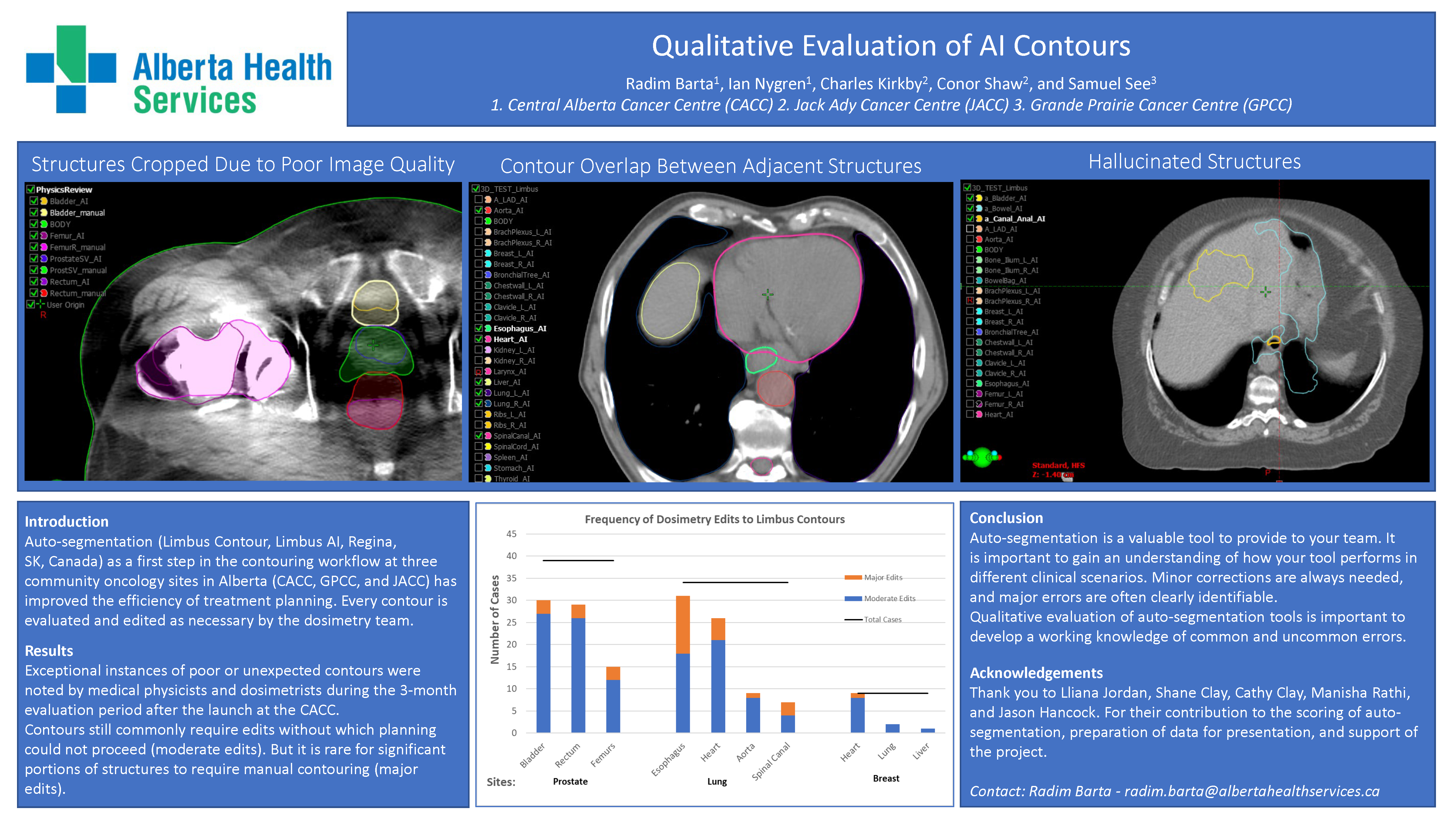
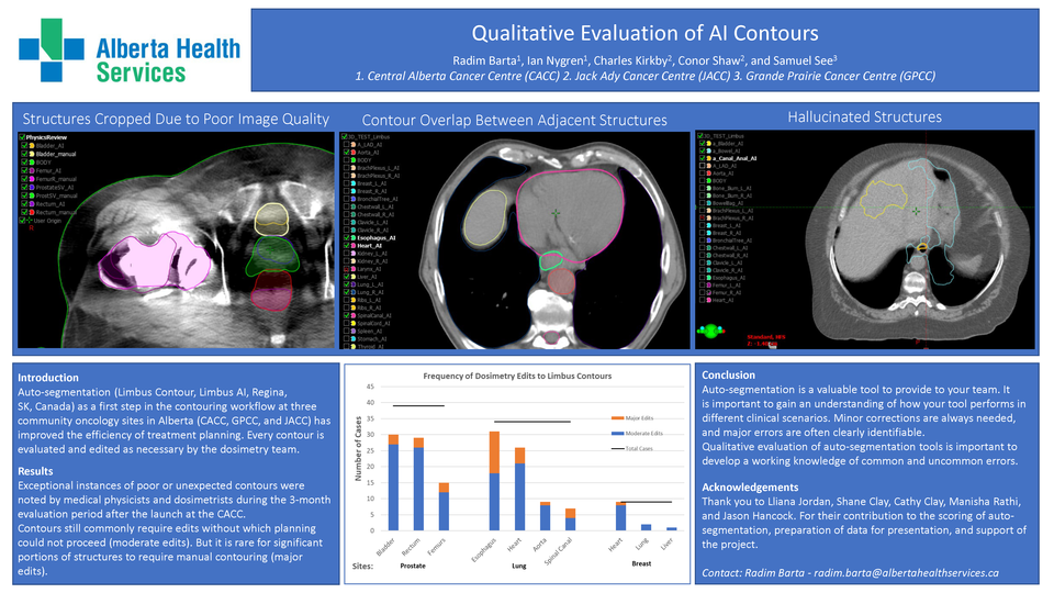
Quantitative measurements are in line with past studies of U-Net algorithm based auto-segmentation. Qualitatively, auto-segmentation contours differ from the clinical contours for different reasons.
The agreement for bladder (DC=97%, DT=4.3 mm) speaks to its relatively consistent shape and position in the pelvic region, as well as soft tissue contrast. Auto-segmentation generates overlapping bladder and prostate, and this may contribute to disagreement in prostate contours (90%, 7.7 mm) when clinically mutual exclusivity is enforced.
Disagreement for the high-contrast femurs (83%, 12.1 mm) results from variation in the number of slices contoured. While auto-segmentation tends to contour the full extent of the femoral heads, expert-generated contours may truncate to only the relevant slices, i.e. those likely to receive doses relevant for plan optimization or assessment. This was also seen in the rectum (80%, 14 mm) and bowel bag (56%, 33 mm).
Penile bulb (59%, 6 mm) highlights a common evaluation challenge with small structures. Even single slice differences in contours lead to significant DC differences, the 6 mm DT better captures the variation seen.
Conclusions:
This study suggests auto segmentation can be used effectively to begin the contouring process. It allows trained experts to focus more time on difficult contours and defining problem areas.