Prospective Comparison of Late 3T Magnetic Resonance Imaging with Conventional Angiography in Evaluating the Patency of Cerebral AVMs Treated with Stereotactic Radiosurgery
Abstract
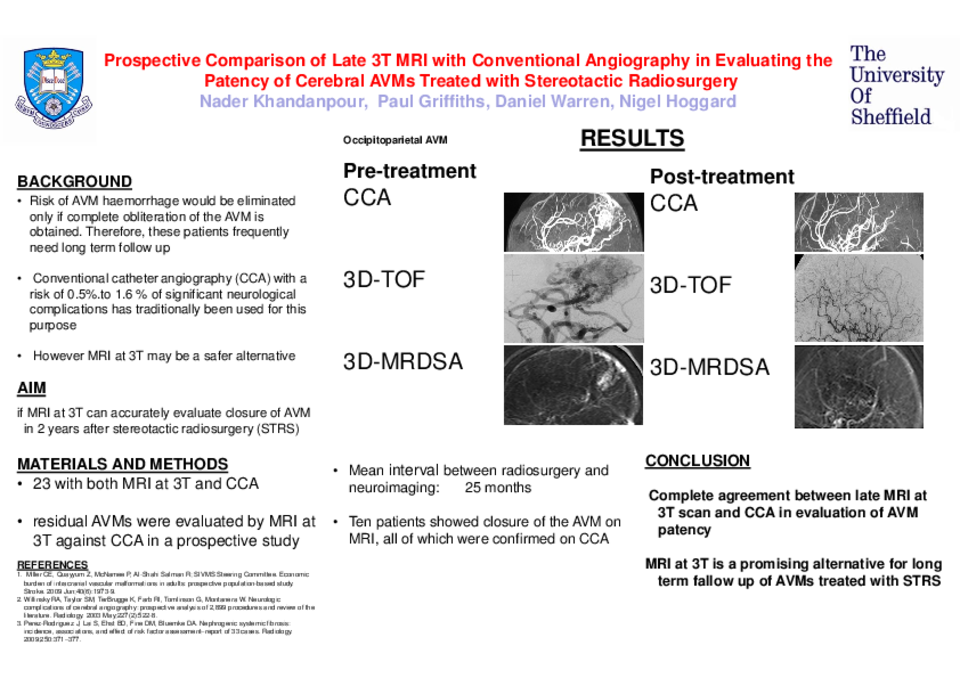
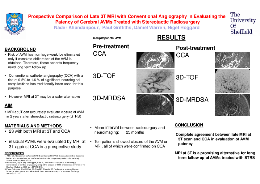
OBJECTIVES: Risk of further haemorrhage in patients suffering from arteriovenous malformation (AVM) would be eliminated only if complete obliteration of the AVM is obtained. Therefore, these patients frequently need long term follow up. Conventional catheter angiography (CCA) with a risk of 0.5%.to 1.6 % of significant neurological complications has traditionally been used for this purpose. However, Magnetic Resonance Imaging (MRI) at 3T may be a safer alternative. The aim of this study was to evaluate if MRI at 3T can accurately evaluate closure of AVM in 2 years after stereotactic radiosurgery (STRS).
METHODS: Twenty three patients with both MRI at 3T and a CCA study. The residual AVMs were evaluated by MRI at 3T against CCA in a prospective study.
RESULTS: The time interval between radiosurgery and neuroimaging was on average of 25 months (range, 15-30 months) for MRI study and 33 months (range, 25-46 months) for CCA study. Ten patients showed closure of the AVM on MRI, all of which were confirmed on CCA.
CONCLUSION: There was a complete agreement between late MRI at 3T scan and CCA in evaluation of AVM patency.
Advances in knowledge: MRI at 3T is a promising alternative for long term fallow up of AVMs treated with STRS.
Related articles