Abstract
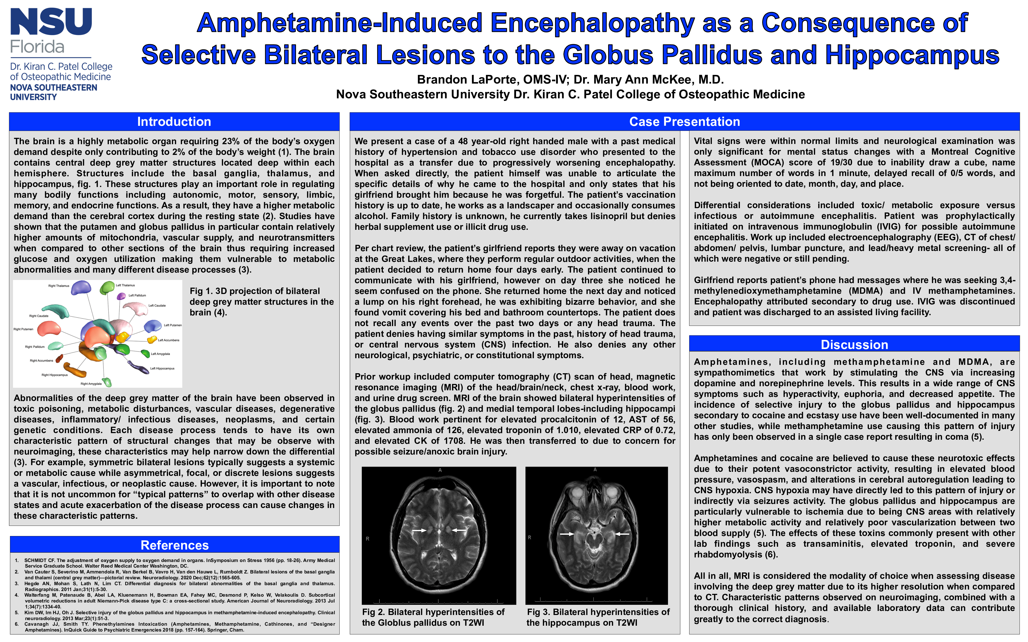
Introduction: The brain contains central deep grey matter structures located deep within each hemisphere. Selective injury of these structures have been observed in toxic poisoning, metabolic disturbances, vascular diseases, degenerative diseases, inflammatory/ infectious diseases, and neoplasms. Each disease process tends to have its own characteristic pattern of structural changes that may be observe with neuroimaging, these patterns may help narrow down the differential diagnosis. It is important to note that it is not uncommon for “typical patterns” to overlap with other disease states and acute exacerbation of certain disease processes can cause changes in these characteristic patterns. The incidence of selective injury to the globus pallidus and hippocampus secondary to cocaine or ecstasy use have been well-documented in many studies, while Kim et al. submitted the only case report of methamphetamine use attributing to this pattern of injury, resulting in coma.
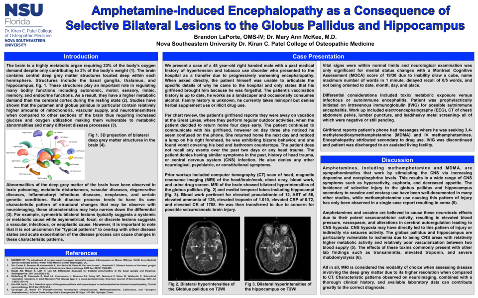
Case Description: We present a case of a 48 year-old right handed male who presented to the hospital as a transfer due to progressively worsening encephalopathy. Per chart review, the patient’s girlfriend reports that the patient returned home early while they were on vacation, however after three days she noticed he seem confused while talking on the phone. Upon returning home, she noticed a lump on his right forehead, exhibition of bizarre behavior, and vomit covering their bed and bathroom countertops. The patient does not recall any events over the past two days or head trauma. Prior workup was notable for an MRI of the brain showing bilateral hyperintensities of the globus pallidus and medial temporal lobes- including hippocampi. Blood work pertinent for procalcitonin of 12 ng/mL, AST of 56 IU/L, ammonia of 126 Umol/L, troponin of 1.010 ng/mL, CRP of 0.72 mg/dL, and CK of 1708 U/L. He was then transferred due to concern for possible seizure/anoxic brain injury. Vital signs were within normal limits and neurological examination was only significant for mental status changes with a Montreal Cognitive Assessment score of 19/30. Differential considerations included toxic/ metabolic exposure versus infectious/ autoimmune encephalitis. Patient was prophylactically initiated on intravenous immunoglobulin and work up included electroencephalography, CT of chest/ abdomen/ pelvis, lumbar puncture, and lead/heavy metal screening. The patient’s girlfriend reports that the patient’s phone had messages where he was seeking ecstasy and intravenous methamphetamines. Encephalopathy attributed secondary to drug use. IVIG was discontinued and patient was discharged to an assisted living facility
Discussion: Determining the cause of deep grey matter lesions can be difficult. However, this case illustrates that certain characteristic patterns observed on neuroimaging, combined with a thorough clinical history, and available laboratory data can contribute greatly to the correct diagnosis.