Abstract
Introduction: Terminally located on the cellular glycocalyx, Sialic acid residues are the hallmark of healthy cells. Malignant transformation of lung tissue disrupts sialylations and sialic acid modifications, delivering a biomarker for neoplasms detection. The alterations are associated with abnormal expression of carbohydrate ligands of adenocarcinomas. We hypothesize that altered sialyl residues in cancerous lung tissues will be detectable with Trithrichomonas mobiliensis lectin (TML). The localized sialyl residues will be digitally assessed in histological sections.
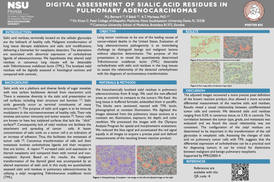
Methods: We histochemically localized sialyl residues in pulmonary adenocarcinomas from 8 lungs. The non-affected areas were used as controls. Each lung tissue was fixed, processed with paraffin, sectioned, reacted with the TML lectin then examined. Sections were digitized at constant set, illumination, exposure, bit depth, and color rendition, the density for the resulting brown product was measured. Images were edited and processed with the Olympus Analysis Program for more precise pixel definition.
Outcomes: Results reveal a causal relationship between undifferentiated tumor cells and controls. We detected sialic acid residues ranging from 0.5% in cancerous tissue to 3.3% in controls.
Discussion: Assessing the changes of sialic acid on pulmonary tumor cells with TML suggests that the differential expression of carbohydrates can be a practical tool for diagnosing tumors. It can be critical for distinctions between malignant and benign pulmonary neoplasms. Supported by PFRG2003-4


