Abstract
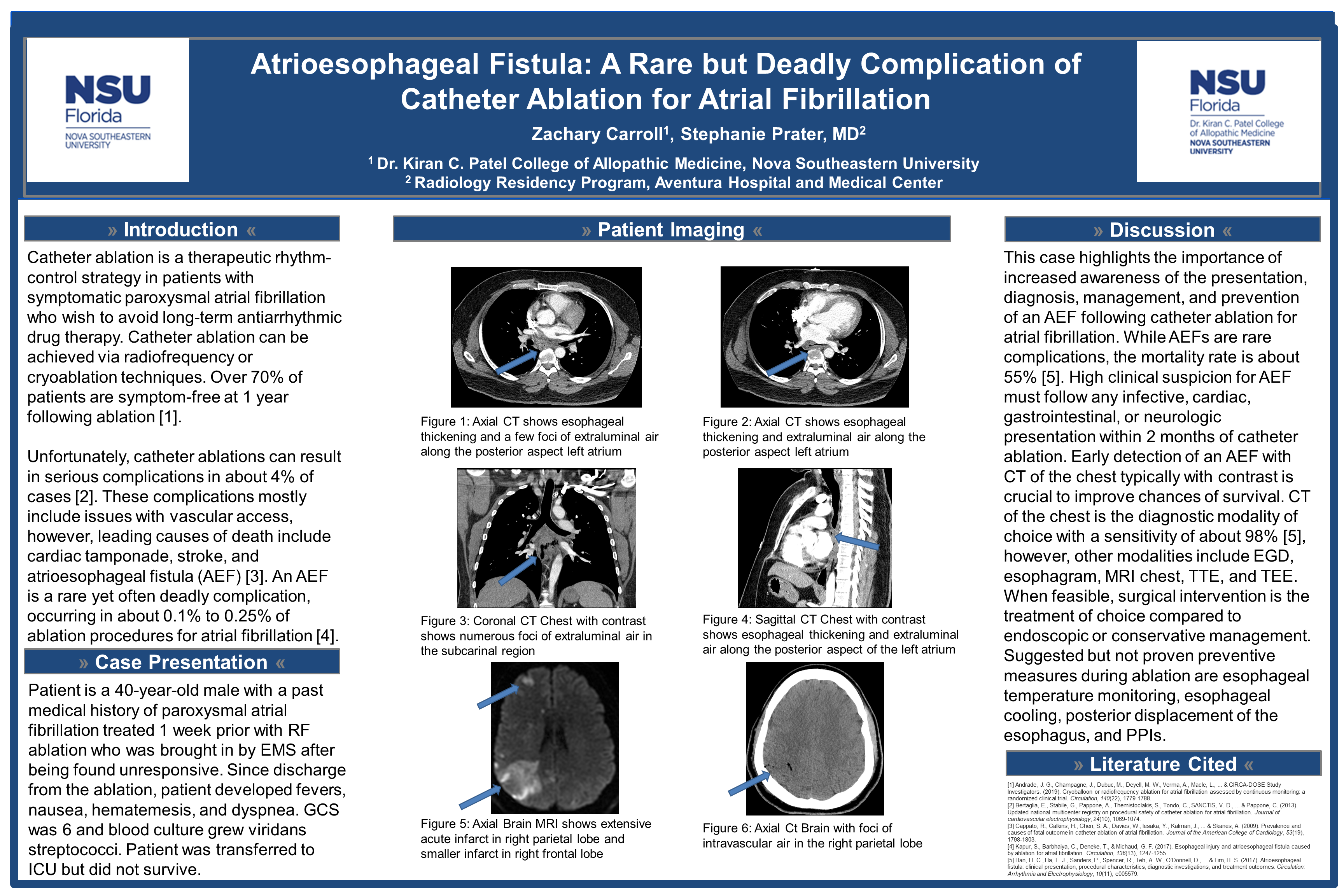
We present a case of a 40-year-old male with a past medical history of paroxysmal atrial fibrillation brought into the emergency room by EMS after being found unconscious. Earlier in the week prior to admission, the patient underwent a planned radiofrequency ablation of an ectopic focus in the pulmonary vein. CT of the chest was suggestive of an atrioesophageal fistula and CT of the brain revealed several foci of air within the brain. Further workup with MRI of the brain showed multifocal infarcts consistent with a stroke due to air emboli that originated from the patient’s atrioesophageal fistula. The patient did not survive. This case highlights the importance of understanding the potential complications of catheter ablation for the treatment of atrial fibrillation. An atrioesophageal fistula is a rare yet often deadly complication, occurring in about 0.1% to 0.25% of ablation procedures for atrial fibrillation. This case also emphasizes the role of imaging in the diagnostic workup of these complications.