Do Conventional Computed Tomography Characteristics of Pathological Neck Nodes Apply after Chemoradiation?
Abstract
Background and purpose:
Computed tomography (CT) continues to be at the forefront in evaluating residual disease after chemoradiation (CRT), however, no definitive CT characteristics have yet produced an effective prognosticator to pathological disease. The purpose of this study was to identify CT characteristics that can properly imply pathologically positive lymph nodes in patients treated with CRT.
Materials and Methods
This study included 91 patients with node-positive head and neck squamous cell carcinoma treated with CRT. All patients had a pre CRT CT and a CT 6-8 weeks post CRT followed by a neck dissection. CT characteristics such as extracapsular extension, heterogeneity, greatest axial dimension, nodal volume and nodal regression were documented and correlated to neck dissection outcomes.
Results:
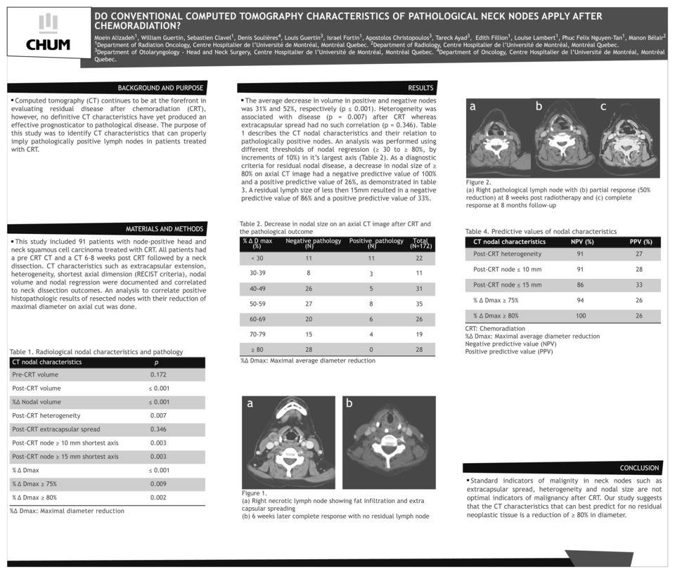
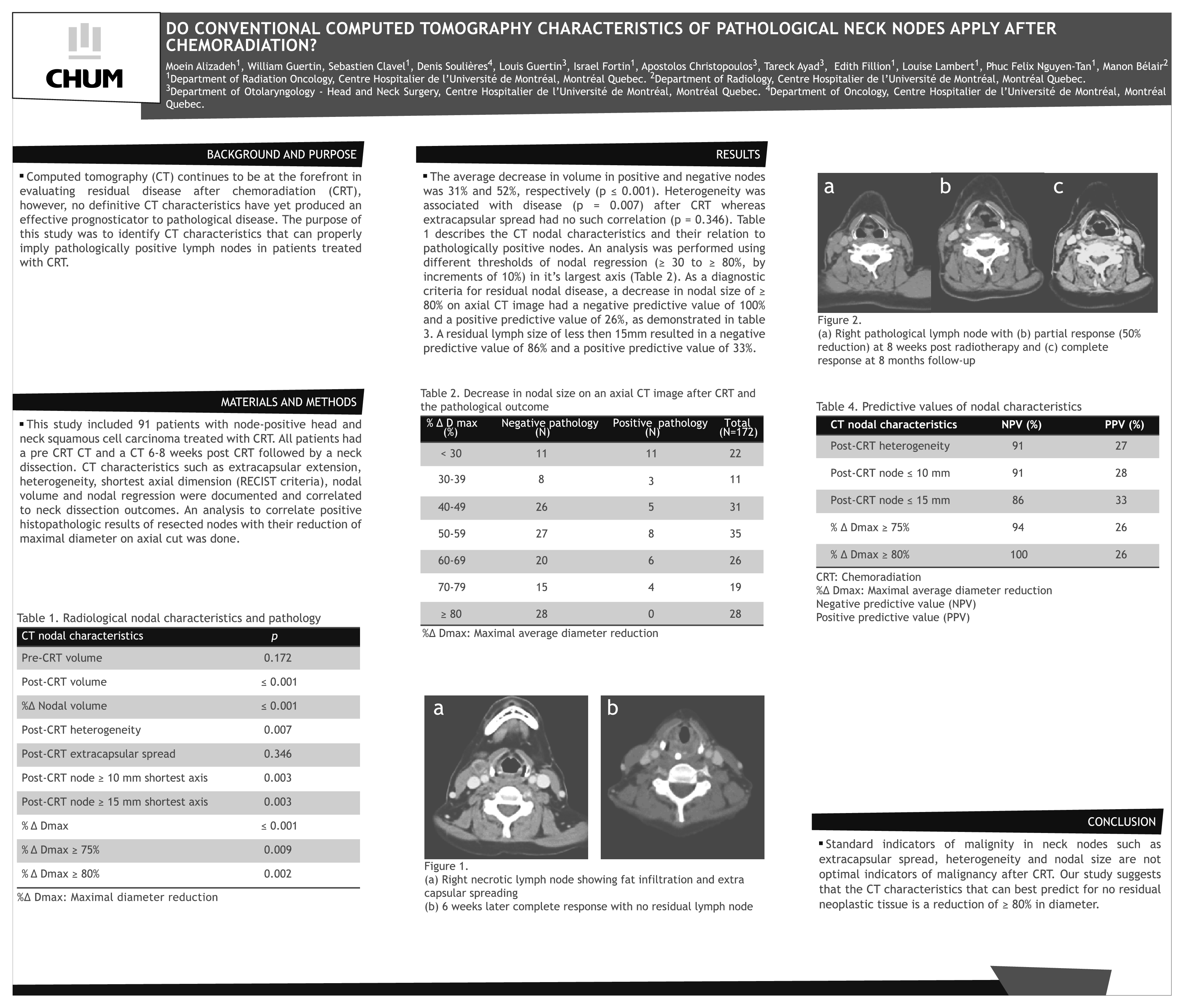
The average decrease in volume in positive and negative nodes was 31% and 52%, respectively (p ≤ 0.001). Heterogeneity was associated with disease (p = 0.007) after CRT whereas extracapsular spread had no such correlation (p = 0.346). A decrease in nodal size on an axial CT image ≥ 80% had a in a negative predictive value of 100% and a positive predictive value of 26%. A residual lymph size of less then 15mm resulted in a negative predictive value of 86% and a positive predictive value of 33%.
Conclusions:
Standard indicators of malignity in neck nodes such as extracapsular spread, heterogeneity and nodal size are not optimal indicators of malignancy after CRT. Our study suggests that the CT characteristics that can best predict for no residual neoplastic tissue is a reduction of ≥ 80% in diameter.
Related articles