Abstract
Introduction and Objectives: Up to 30% of low birth weight preterm infants manifest some form of periventricular white matter injury (PWMI) making it the most common form of brain injury affecting premature infants. It is believed that loss of oligodendrocyte progenitor cells (OPCs), which are proliferative cells that develop into myelinating OLs, plays a major role in PWMI causation. Presently, few pharmacological approaches specifically target OPCs resulting in increased proliferation of these cells and increased brain myelination. We discovered that diazoxide (DZ), an activator of ATP-sensitive potassium channels (KATP), promotes OPC proliferation and attenuated hypoxia induced brain injury in neonatal mice. Out of 610 Diazoxide derivatives, compound K261-0298 (ChemDiv) was identified as the most potent stimulator of myelination among our lead compounds.
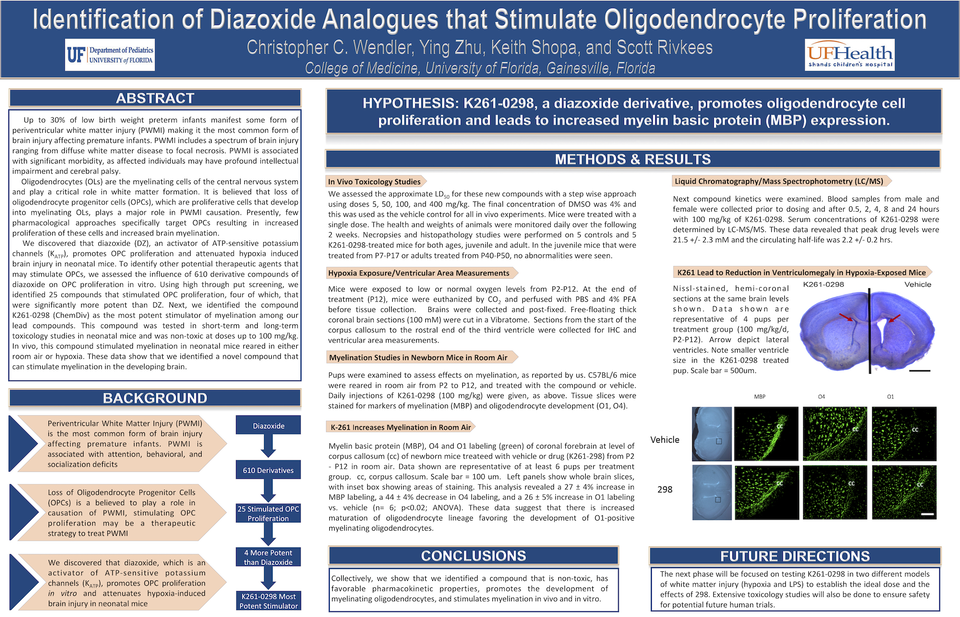
Methods: Using in vivo toxicology studies we assessed the approximate LD50 for K261-0298 in a stepwise approach. Next compound kinetics were examined via liquid chromatography/mass spectrophotometry (LC/MS) on mice blood samples. Myelination studies in newborn mice reared in room air were performed to determine markers of myelination and oligodendrocyte proliferation. Hypoxia exposure with follow-up ventricular area measurements were conducted to determine effect on ventriculomegaly in hypoxia exposed mice.
Results: In the juvenile mice that were treated from P7-P17 oradults treated from P40-P50, no abnormalities were seen. Blood samples from male and female were collected prior to dosing and after 0.5, 2, 4, 8 and 24 hours with 100 mg/kg of K261-0298. Data from serum LC-MS/MS revealed that peak drug levels were 21.5 +/- 2.3 M and the circulating half-life was 2.2 +/- 0.2 hrs. Tissue slices were stained for markers of myelination (MBP) and oligodendrocyte development (O1, O4). This analysis revealed a 27 ± 4% increase in MBP labeling, a 44 ± 4% decrease in O4 labeling, and a 26 ± 5% increase in O1 labeling vs. vehicle (n=6; p<0.02; ANOVA). These data suggest that there is increased maturation of oligodendrocyte lineage favoring the development of O1-positive myelinating oligodendrocytes. At the end of the treatment period mice were examined for ventriculomegaly, as reported. We observed a marked reduction in ventriculomegaly in the K261-0298 (0.0054 +/- 0.00013 m2) vs. vehicle-treated (0.0156 +/- 0.0039 m2) mice (n= 4) per treatment, p<0.03).
Conclusions-Implications: Collectively, we show that we identified a compound that is non-toxic, has favorable pharmacokinetic properties, promotes the development of myelinating oligodendrocytes, and stimulates myelination in vivo and in vitro. The next phase will be focused on testing K261-0298 in two different models of white matter injury (hypoxia and LPS) to establish the ideal dose and the effects of 298. Extensive toxicology studies will also be done to ensure safety for potential future human trials.