Violet, an Antibody to Ventricular Progenitor Cells
Abstract
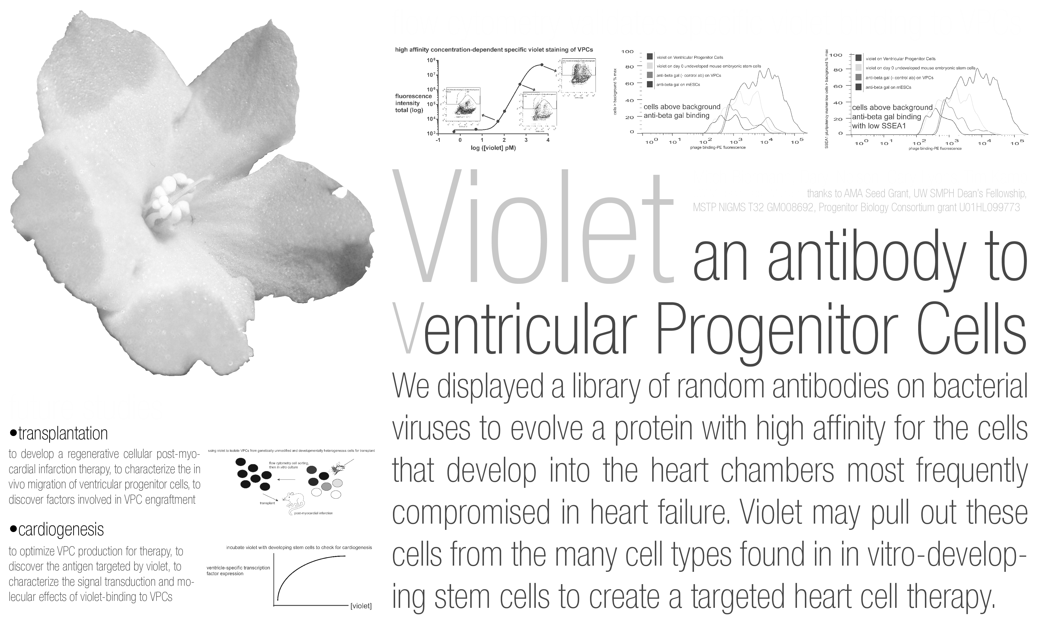
We have created a monoclonal antibody that specifically binds ventricular progenitor cells, the developmental parent cells of the myocardium most commonly damaged in heart disease. We will use it to obtain VPCs from unmodified developing stem cells for transplant.
BACKGROUND: The death of ventricular cardiomyocytes is the basis of the most severe forms of heart disease such as myocardial infarction. Because the heart exhibits limited repair potential, stem cell transplantation is being investigated to repair injured myocardium. Given the demonstrated limitations of using either fully pluripotent stem cells or fully differentiated cardiomyocytes, one hypothesis is that an intermediate ventricular progenitor cell (VPC) may be ideal for transplantation. VPCs have the potential to develop into the cardiomyocytes most commonly damaged by heart disease and are intermediate in development compared to cells previously used in animal transplantation studies. However, no unique VPC surface markers exist, and so it is impossible to isolate VPCs from the many lineages found in unmodified developing stem cells without genetically altering the cells to insert foreign transgene reporter systems, an undesirable feature for therapeutic cells.
METHODS: We used phage display to pan a library of random antibodies against a line of mouse embryonic stem cells (mESCs) expressing hygromycin resistance in a subset of cells previously shown to develop a predominantly ventricular cardiomyocyte fate. Hygromycin-derived VPCs were then mixed with mESCs and our antibody library, and fluorescence-activated cell sorting was used with SSEA1 (an mESC marker) to select phages attached only to VPCs.
RESULTS: A monoclonal antibody (Violet) was shown in flow cytometry to bind VPCs with higher affinity than a phage negative control antibody, and Violet bound a greater proportion of VPCs than mESCs. The parent polyclonal antibody mixture and five other monoclonals were less or not at all specific for VPCs, respectively. DNA sequencing revealed Violet represented 10/16 antibodies in the mixture. These data suggested that Violet was a tight-binding antibody to VPCs, which was confirmed by dose response analysis showing concentration-dependent Violet binding of VPCs at nanomolar concentrations. Violet preferentially labeled VPCs with low expression of the pluripotency marker SSEA1.
CONCLUSIONS: We have identified a monoclonal antibody that specifically binds VPCs. As Violet labels an enriched SSEA1- subset of VPCs, in the future we will characterize the differentiation fate of these SSEA1- cells, and we will use Violet to obtain VPCs from unmodified developing mESCs for transplantation studies in a mouse model of myocardial infarction.
Related articles