Abstract
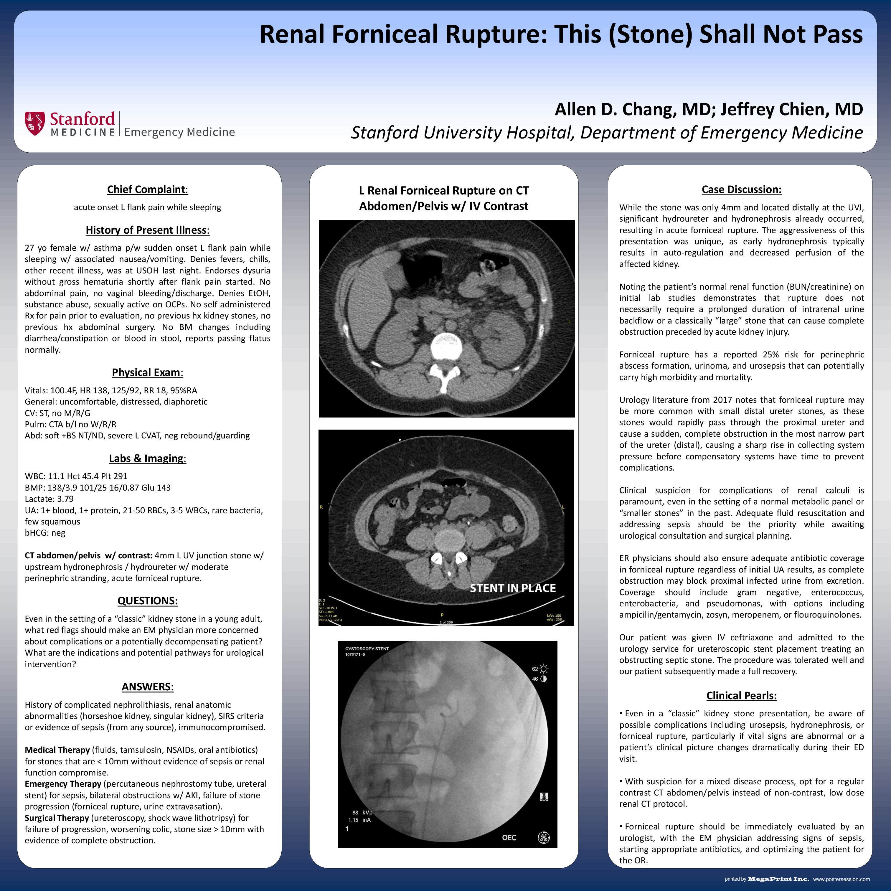
Forniceal rupture has a reported 25% risk for perinephric abscess formation, urinoma, and urosepsis that can potentially carry high morbidity and mortality.
Urology literature from 2017 notes that forniceal rupture may be more common with small distal ureter stones, as these stones would rapidly pass through the proximal ureter and cause a sudden, complete obstruction in the most narrow part of the distal ureter, causing a sharp rise in collecting system pressure before compensatory systems have time to prevent complications.
Clinical suspicion for complications of renal calculi is paramount, even in the setting of a normal metabolic panel or “smaller stones” in the past medical history. Adequate fluid resuscitation and addressing signs of sepsis should be the priority while awaiting urological consultation and surgical planning.
The culprit stone in this case was only 4mm and located distally at the UVJ. Significant hydroureter and hydronephrosis was already present, resulting in acute forniceal rupture. The aggressiveness of this presentation was unique, as early hydronephrosis typically results in auto-regulation and decreased perfusion of the affected kidney.
Noting the patient’s normal renal function (BUN/creatinine) on initial lab studies demonstrates that rupture does not necessarily require a prolonged duration of intrarenal urine backflow or a previously classically “large” stone to cause simultaneous kidney injury and complete urinary obstruction.
ER physicians should ensure adequate antibiotic coverage in forniceal rupture regardless of initial UA results, as complete obstruction may block proximal infected urine from excretion. Coverage should include gram negative, enterococcus, enterobacteria, and pseudomonas, with options including ampicilin/gentamycin, pipercillin-tazobactam, meropenem, or flouroquinolones.
• Even in a “classic” kidney stone presentation, be aware of possible complications including urosepsis, hydronephrosis, or forniceal rupture, particularly if vital signs are abnormal or a patient’s clinical picture changes dramatically during their ED visit.
• With suspicion for a mixed disease process, opt for a regular contrast CT abdomen/pelvis instead of non-contrast, low dose renal CT protocol.
• Forniceal rupture should be immediately evaluated by an urologist, with the EM physician addressing signs of sepsis, starting appropriate antibiotics, and optimizing the patient for the OR.