Abstract
Background
Regarding surface high-dose rate (HDR) brachytherapy, the status quo approach to positioning catheters is the Freiburg flap, which is limited with regard to conformity to the surface anatomy and the presence of air between the spheres. Three-dimensional (3D) printing is suitable for the fabrication of complex customized structures. The objective of this study is to illustrate through a patient case the application of 3D printing for surface HDR brachytherapy, namely to create a conformal personalized surface applicator.
Methods
To treat a squamous cell carcinoma in the left supra-alar region of the nose, an applicator for surface brachytherapy was 3D-printed. For the design process, a CT dataset of the patient’s head was imported in the treatment planning system (Eclipse 10, Varian Medical Systems). A 10 mm thick bolus object was created to surround the PTV by at least 20 mm in all directions and to allow stability and reproducibility of the surface applicator during daily placement. Using methods developed in-house, nine 3.3 mm diameter tunnels for HDR brachytherapy catheters were created, with lateral spacing of 10 mm, by designing cylinders that followed the contours of the posterior surface of the applicator. The tunnels were placed 5 mm from the tissue interface and a subtraction operator was performed to remove intersecting objects. The applicator was printed using thermoplastic polyurethane and a Lulzbot Taz 5 3D printer. The structure’s fit to the anatomy was assessed and a brachytherapy treatment plan featuring an iridium-192 source was generated using the brachytherapy planning system (Oncentra, Nucletron).
Results
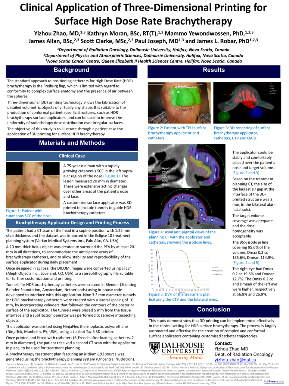
The applicator could be stably placed over the patient’s nose. Based on the treatment planning CT, the size of the largest air gap at the interface of the 3D-printed structure was 2 mm, in the bilateral alar-facial sulci. The treatment plan had adequate coverage and a dose homogeneity acceptable for brachytherapy: 95% isodose line covering 95.6% of the volume, Dmax 0.2 cc 135.6%, Dmean 114.9%.
Conclusions
This study demonstrates the concept that 3D printing can be implemented effectively in the clinical setting for HDR surface brachytherapy. The process is largely automated and effective for the creation of complex and conformal surface applicators containing customized catheter trajectories.


