Abstract
Prostate cancer is typically treated using Image Guided radiation therapy, where a patient is set up onto the treatment bed, the patient’s anatomy is visualized using an imaging modality and the patient’s position is adjusted to match the original radiation treatment plan. Aligning the patient according to the original radiation treatment plan is important for both delivering the correct dose to the target volume as well as for minimizing the radiation doses given to other tissue (and thus side effects).
Previously, the typical imaging modality utilized X-rays. To set patients up consistently, fiducial markers would be surgically implanted into patients beforehand, and the patients would be lined up based on the placement of these markers. These markers must be surgically inserted, cost thousands of dollars to be inserted, and have their inherent risks. With the advent of Cone Beam CT (CBCT) scanners, it is now possible to visualize patient’s soft tissue and thus it may no longer be necessary to insert fiducial markers to align patients before they are treated. There have been three previous studies have had conflicting results on whether there is a significant difference between aligning patients based on fiducial markers versus aligning patients based on their soft tissue. Results of whether fiducial marker versus soft tissue alignment may be site-specific and may depend on the level of training of radiation therapists and their experience with aligning patients based on soft tissue.
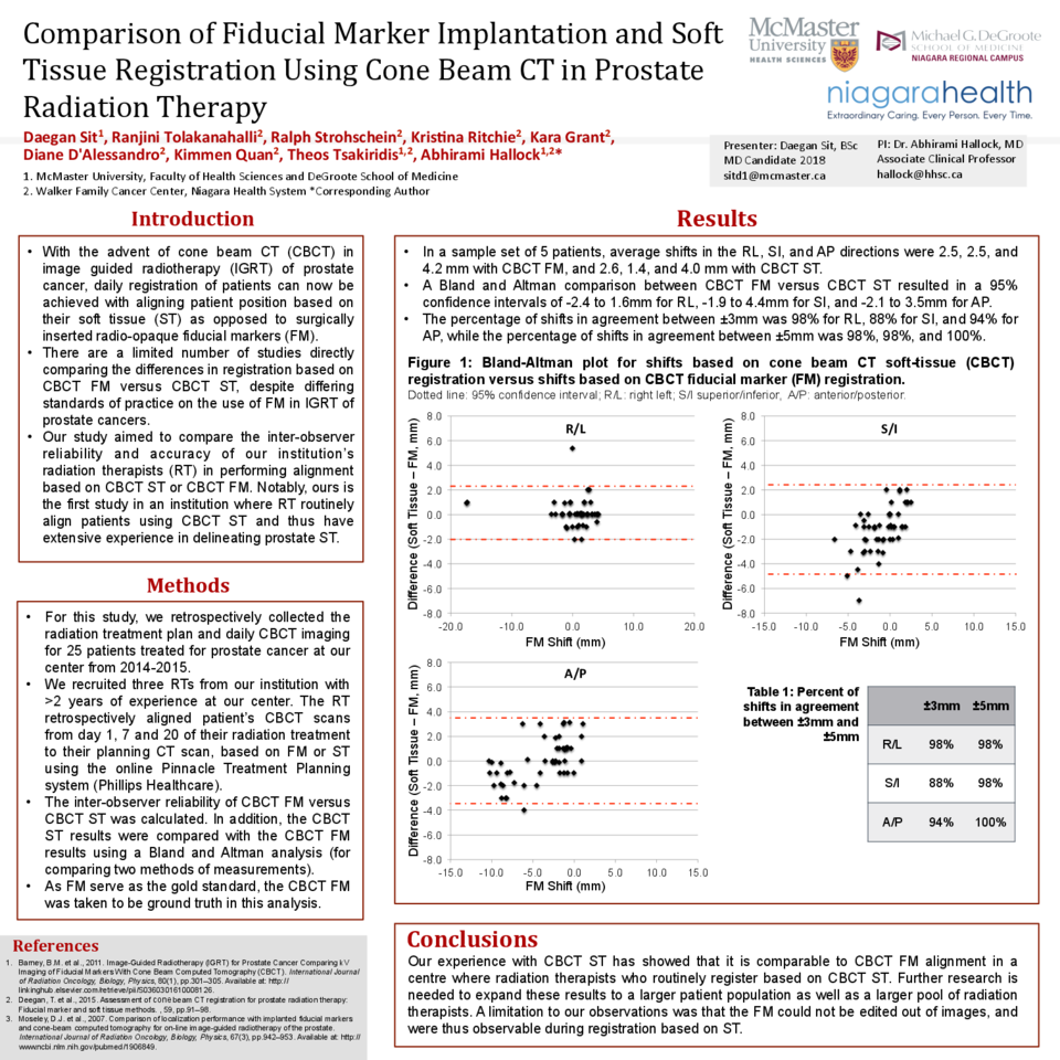
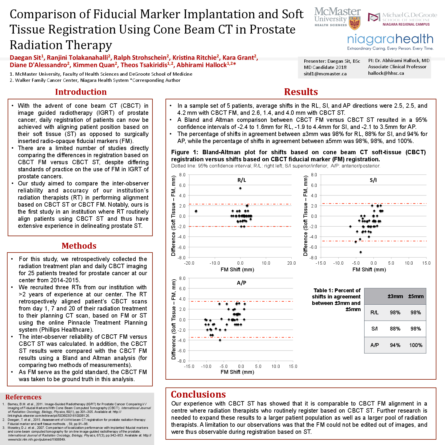
Furthermore, no current studies are representative of a regional cancer care program. This study seeks to compare radiation therapists’ retrospective alignment of patients based on inserted fiducial markers versus the soft tissue of patients. For this study, the CBCT images of 50 patients previously treated at the Walker Family Cancer Center for prostate cancer will be uploaded onto radiation therapy planning software. The radiation therapists will then “shift” the patient images based on either the gold fiducial markers or the patient’s soft tissue. The average differences will then be compared to see if they are significantly different from one another. This study is important as it can help determine if patients undergoing treatment for prostate cancer may skip the insertion of fiducial markers, thus avoiding an invasive procedure and its inherent risks, as well as saving the healthcare system thousands of dollars per procedure.


