Abstract
Purpose
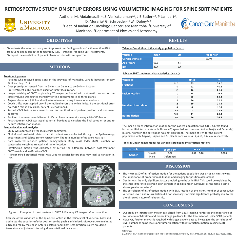
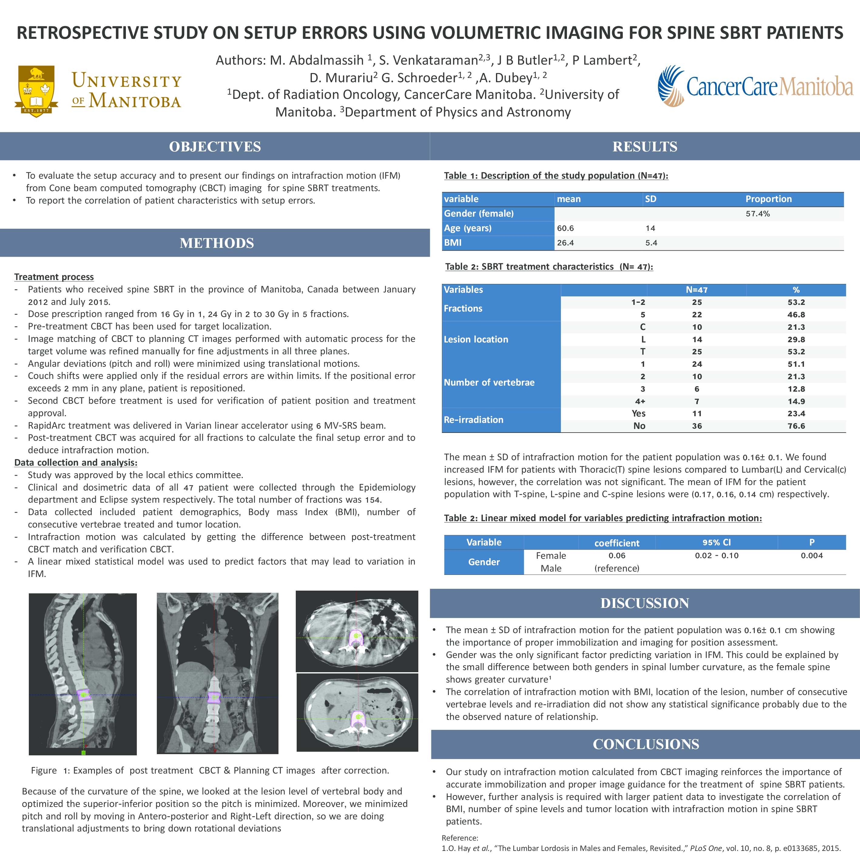
Stereotactic body radiation therapy (SBRT) for spine is challenging due to high-dose gradients sparing the cord in the treatment plans. Proper immobilization and positioning verification with imaging are essential to ensure accurate delivery of radiation. In this study we present our findings of initial setup error and intrafraction motion from Cone beam computed tomography (CBCT) imaging and the correlation of patient characteristics with the above mentioned parameters.
Materials and Methods
Forty-seven spine SBRT patients with a total of 154 fractions treated with the fractionation schedule of 16 Gy in 1, 24 Gy in 2 and 30 Gy in 5 fractions were part of this study. Pre-treatment CBCT was used for localization of the target and couch shifts were applied based on the target volume matching to the planning CT image set. This process included verification of rotational deviations and repositioning of patients if the matching deviation exceeded 2mm. Intrafraction motion (IFM) was calculated by subtracting the couch shifts of initial CBCT from the final CBCT match. RapidArc treatment was delivered in Varian linear accelerator using 6 MV-SRS beam. Post-treatment CBCT was acquired for all fractions to calculate intrafraction motion. Patient characteristics such as Gender, Body mass Index (BMI), number of consecutive vertebrae treated, tumor location were assessed for associations with IFM. A linear mixed statistical model was used to predict factors that may lead to variation in IFM.
Results
The initial setup error calculated from pre-treatment CBCT had a mean of 7.8 mm in consistent with published data. The mean ± SD of intrafraction motion for the patient population was 0.16± 0.1 showing the importance of proper immobilization and imaging for position assessment. Gender was the only significant factor predicting variation in IFM. The correlation of intrafraction motion with BMI, location of the lesion, number of consecutive vertebrae levels and re-irradiation did not show any statistical significance probably due to the the observed nature of relationship. We found increased IFM for patients with Thoracic(T) spine lesions compared to Lumbar(L) and Cervical(c) lesions, however, the correlation was not significant. The mean of IFM for the patient population with T-spine, L-spine and C-spine lesions were (0.17, 0.16, 0.14 cm) respectively.
Conclusions
Our study on intrafraction motion calculated from CBCT imaging reinforces the importance of accurate immobilization and proper image guidance for the treatment of spine SBRT patients. However, further analysis is required with larger patient data to investigate the correlation of BMI, number of spine levels and tumor location with intrafraction motion in spine SBRT patients.


