Novel MRI Techniques for Early Detection of Brain Metastases
Abstract
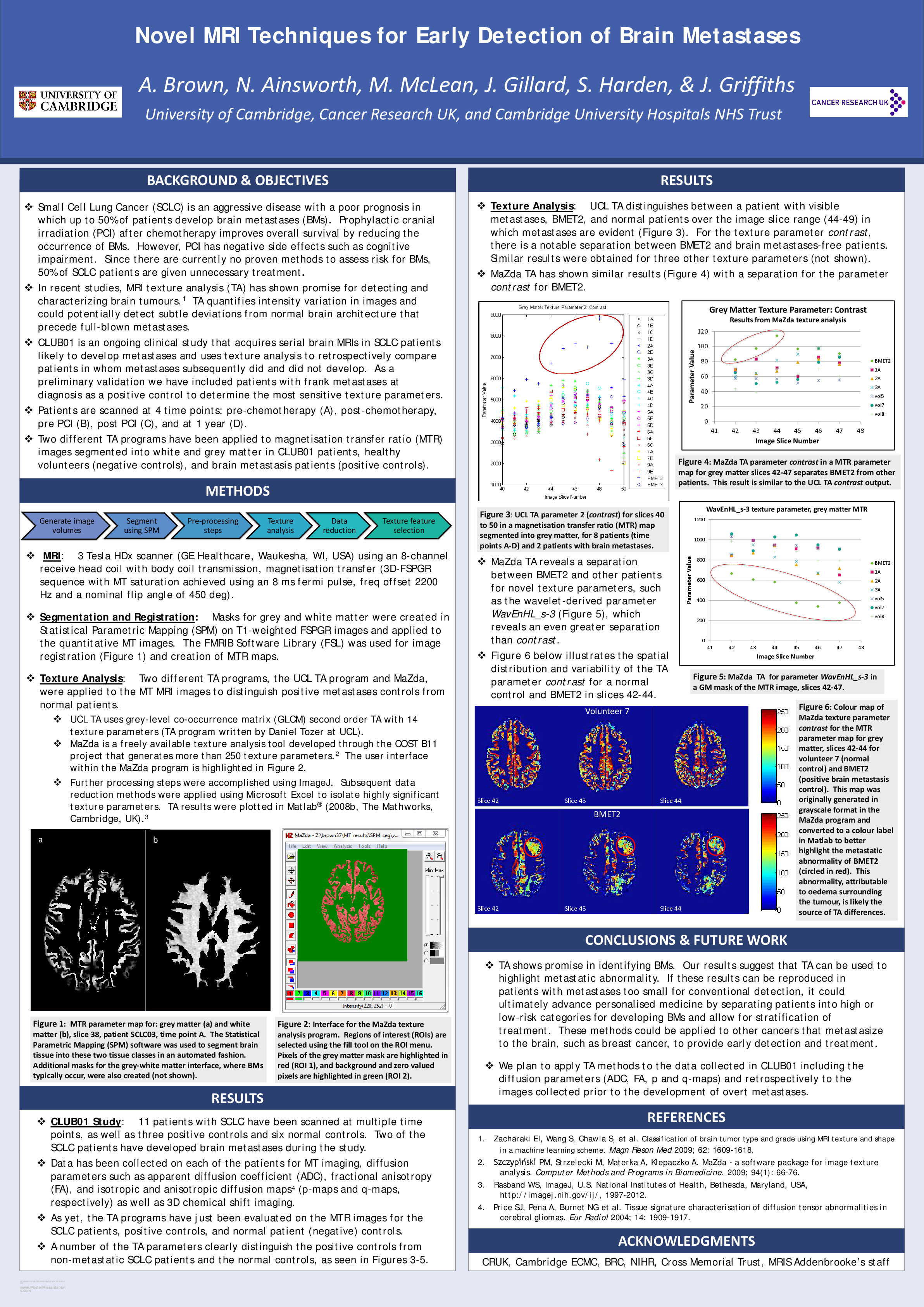
Background and Aims: Small Cell Lung Cancer (SCLC) is an aggressive disease with a poor prognosis in which up to 50% of patients develop brain metastases (BMETs). Prophylactic cranial irradiation (PCI) after chemotherapy improves overall survival by reducing the occurrence of BMETs. However, PCI has negative side effects such as cognitive impairment. Since there are currently no proven methods to assess risk for BMs, 50% of SCLC patients are given unnecessary treatment. In recent studies, MRI texture analysis (TA) has shown promise for detecting and characterizing brain tumours. TA quantifies intensity variation in images and could potentially detect subtle deviations from normal brain architecture that precede full-blown metastases.
The aim of this study is to acquire serial MRI in patients with a high likelihood of developing metastases and retrospectively compare patients in whom metastases subsequently did and did not develop; as a preliminary validation we have included patients with frank metastases at diagnosis as a positive control to see which texture parameters detect metastases most sensitively.
Methods: Experiments were performed on a 3 Tesla HDx scanner (GE Healthcare, Waukesha WI, USA) using an 8-channel receive head coil with body coil transmission. MRI scans included magnetization transfer (3D-FSPGR sequence with MT saturation achieved using 8ms fermi pulse, freq offset 2200Hz and a nominal flip angle of 450 deg) and diffusion-weighted images (EPI with 25 gradient directions and b values of 0 and 1000). Ten patients with SCLC and three patients with brain metastases were scanned. SCLC patients were scanned at four time points, spanning the period from pre-treatment to follow-up at one year. The raw data was processed to create magnetization transfer ratio (MTR) maps as well as apparent diffusion coefficient (ADC), fractional anisotropy (FA), and isotropic and anisotropic diffusion maps (p-maps and q-maps, respectively). In order to better detect abnormal pixel regions, we applied segmentation masks for grey and white matter created in Statistical Parametric Mapping (SPM) on T1-weighted FSPGR images to the quantitative MT and diffusion images. The FMRIB Software Library (FSL) FLIRT tool was used for image registration.
In addition, we have applied grey-level co-occurrence matrix (GLCM) second order TA to MRI images to examine its ability to distinguish positive metastases controls from normal patients for 14 texture parameters (TA program written by Daniel Tozer at UCL). Further processing steps, such as image type conversion and grey-level normalization, were accomplished using ImageJ and TA results were plotted in Matlab® (2008b, The Mathworks, Cambridge, UK).
Results and conclusions: TA distinguishes between a patient with visible metastases, BMET2, and normal tissue during the image slice range (44-49) in which metastases are evident for several texture parameters studied for grey matter magnetization transfer ratio (MTR) maps. For the texture parameter "Contrast," there is a notable separation between BMET2 and brain metastases-free patients in a clinical study. We have obtained similar results with three other texture parameters.
Overall, we conclude that TA shows promise in identifying BMETs. Our results suggest that TA can be used to highlight metastatic abnormality. If these results can be reproduced in patients with metastases too small for conventional detection, it could ultimately advance personalized medicine by separating patients into high or low-risk categories for developing BMs and allow for stratification of treatment. These methods could be applied to other cancers that metastasize to the brain, such as breast cancer.
Related articles