Abstract
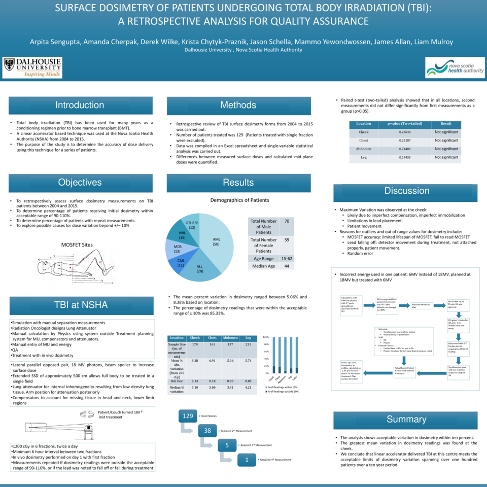
Purpose: Total body irradiation (TBI) is used prior to bone marrow transplantation as part of the conditioning regimen in selected patients. A linear accelerator based technique has been used at our treatment centre, between 2004 and 2015. Compensators to account for missing tissue in the head and neck and lower leg regions, as well as a lung attenuator for internal inhomogeneity resulting from low density lung tissue are routinely used. Dose variation within ten percent of the prescribed midplane dose is considered acceptable. The purpose of this study was to determine whether dose variation was within acceptable limits for patients who underwent TBI.
Materials and Methods: Following chart review, 129 patients between June 2004 and August 2015 who received TBI in six fractions were included in this study. Patients receiving single fraction treatment were excluded. MOSFET dosimetry was used to measure surface dose at 4 or 5 locations when patients received the first fraction of TBI. Dosimetry was repeated during the second fraction for any site with variation greater than ten percent, or when MOSFET position was noted to have shifted. Statistical analysis on patient data, diagnosis and dosimetry measurements was carried out using a Microsoft Excel spreadsheet.
Results: Of the 129 patients who met the inclusion criteria, 50 were diagnosed with AML, 30 with ALL and 11 with CML. The rest of the patients were diagnosed with lymphoma or MDS. The mean percent variation in dosimetry ranged between 3.5% and 8.3%. The highest variation was found in cheek dosimetry. A high percentage of dosimetry readings (85.5%) were within the acceptable range. The highest number of individual readings outside ± 10% was found at the leg. The median percentage variation was low (3.3% to 5.1%) depending on location.
Conclusion: A retrospective analysis of 129 patients was carried out for the period 2004 to 2015. The analysis shows acceptable variation in dosimetry within ten percent. The top three locations with greatest variation were the cheek, the chest, and the leg respectively. We conclude that linear accelerator delivered TBI at our centre meets the acceptable limits of dose variation for 129 patients over a ten-year period. The reasons for variation at particular sites will be discussed.


