Abstract
Testicular germ cell tumours (TGCT) are the most common malignancy in males aged 20 to 39, and increasing in incidence. TGCTs have a tendency to grow rapidly and have a high risk of metastatic spread. TGCTs commonly present with a palpable mass, yet may present with symptoms arising from metastatic disease. TGCTs have a propensity to metastasis to retroperitoneal lymph nodes, lungs, liver, bones and less frequently to the brain. The phenomenon of a primary TGCT outgrowing its blood supply and undergoing auto-infarction has been described as a “burned out” TGCT. Despite initial diagnosis with metastatic disease, therapeutic strategies for the treatment of TGCTs represents a paradigm shift to curable disease.
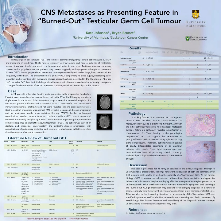
A 24 year-old otherwise healthy male presented with progressive headaches. Initial CT and MRI imaging reported a single mass in the frontal lobe. Complete surgical resection revealed suspicion for metastatic poorly differentiated carcinoma with an inconclusive immunohistochemical profile. CT and PET scans revealed pulmonary and osseous metastases. Follow up MRI revealed intracranial disease progression and he underwent whole brain radiation therapy (WBRT). Additional testing with chromosomal analysis revealed features consistent with a TGCT. Scrotal ultrasound revealed a minimally atrophic right testis. With evidence supporting the potential for complete response to chemotherapeutic treatment in GCT, the patient was started on cisplatin and etoposide. Unfortunately, the patients rapidly deteriorated before receiving full course of treatment.
A salient feature of all invasive TGCTs is a gain in material from the short arm of chromosome 12, and is diagnostic if present. Although the initial pathology revealed a non-diagnostic metastatic tumour, further testing revealed amplification of chromosome 12p leading to the diagnosis of TGCT. This suggests that examination of poorly differentiated carcinomas of an unknown primary site using light microscopy and immunohistochemical profiling alone may be inadequate, and should undergo molecular chromosomal analysis.
This case is presented for its unconventional presentation, rarity of occurrence. It brings forward the discussion of both the commonality of GCT in young male adults, as well as the anomaly of a “burned out” GCT. With unreliable tumour markers, nonspecific symptoms and pathological findings, the ‘burned out’ GCT phenomenon may account for challenging diagnosis in a variety of cases, especially with the presenting symptom arising from a less common metastatic site. This case adds to the increasing literature on a rare entity of the “burned-out” GCT, and upon literature review, presents itself as the first reported case presenting with brain metastasis. By establishing a firm base of literature and a familiarity of the diagnostic process, a deeper understanding into medical management may arise.